
Consumer tech has got very good at rearranging tiny packets of energy. Your phone steers radio waves around crowded cities, earbuds beam audio through skin and bone, and a smartwatch uses milliamps of current to build a picture of your heartbeat from faint changes in light.
Medical devices do a similar kind of work, but with a more direct target: human tissue. Instead of drawing a map on a screen, the aim is often to write a pattern into the body itself, with enough precision that the tissue then repairs or reorganizes on cue.
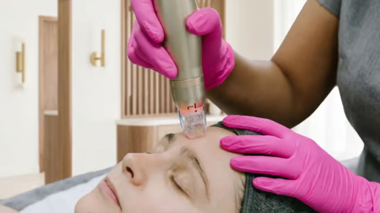
Radiofrequency microneedling sits squarely in that category. It looks like a cosmetic gadget, a handpiece with a throwaway cartridge of pins, but inside it is closer to a controlled welding rig for the skin, calibrated down to fractions of a millimeter and a few degrees Celsius.
From Handset to Skin: A Guided Energy Delivery System
A typical RF microneedling device combines three subsystems.
- A motorized or spring-loaded needle array
- A radiofrequency generator and matching circuit
- A control unit that coordinates timing, depth, and power
The cartridge at the tip carries a grid of very fine needles, often 10 to 64 pins. They are usually stainless steel or gold plated, with diameters in the order of a human hair. Some are insulated along the shaft so that only the tips conduct; others are uninsulated for a different heat profile.
When the operator triggers a pulse, the device drives the needles into the skin to a pre-set depth, anywhere from around 0.5 millimeters to 4 millimeters. At full extension, the tips sit in the dermis, the layer of skin where collagen fibers, blood vessels, and other support structures live.
Only once the pins are in place does the radiofrequency circuit fire.
The handset connects those pins to an RF generator similar in principle to the unit in a small radio transmitter or diathermy device. It produces an alternating current at a specific frequency, typically in the several hundred kilohertz to a few megahertz range, and pushes it through the array for a few tens or hundreds of milliseconds.
Skin does not absorb radiofrequency like an antenna absorbs a Wi-Fi signal. At these settings, RF is mainly a way of delivering current into a resistive material. The current meets electrical resistance as it moves through tissue, and that resistance converts electrical energy into heat.
The interesting detail is where that heat appears and how sharply it is confined.
Fractional Arrays: Making Microscopic Hot Pixels
Older RF skin devices used surface electrodes. The current ran between plates or across a broad tip and created a wide zone of heating. Microneedling devices replace that broad contact with an array of tiny, spaced conductors poked directly into the dermis.
Each needle tip behaves like a local electrode. When the RF pulse fires, current leaves a tip, flows through the surrounding tissue, and returns through another part of the circuit. Because the conducting point is small and the path is short, the current density near the tip is high. That is where the most heat is produced.
Thermally, the pattern looks like a grid of three-dimensional pixels. Around each tip, a small column or ovoid of tissue is heated to temperatures high enough to cause controlled injury, often in the range of roughly 60 to 70 degrees Celsius, while the tissue between pins sits cooler and intact.
This is why you will often see the treatment described as fractional. Only a fraction of the surface area is directly heated at any one pass, in the same way that a fractional laser draws a dotted matrix of microscopic burns rather than sweeping the whole area in a solid block.
This spatial pattern does two main things:
- It allows fairly aggressive local heating without destroying the entire sheet of skin.
- It keeps “bridges” of untouched tissue between columns, which speeds repair.
All of this is governed by the device’s geometry and timing. Pin diameter and spacing set the basic grid. Pulse duration and power set how much heat accumulates around each tip before it can diffuse away.
How Depth and Energy Are Controlled
On the surface, a user interface might offer simple options: depth in millimeters and intensity on a numbered scale. Underneath, the device is juggling mechanics and electronics.
Mechanical Depth Control
Depth is controlled by the throw of the needle actuator. In a common design, the cartridge sits on a cam or linear motor. When activated, the mechanism drives the pins out to a set position, then retracts them when the pulse is over.
The control unit reads or sets this position through step counts in a motor, magnetic sensors, or mechanical stops. Change the depth on the screen and the device changes how far the pins extend from the cartridge.
A few millimeters sounds trivial, but in skin it matters. The epidermis, the outer layer, is usually under 0.2 millimeters thick on the face. The dermis below ranges from roughly 1 to 4 millimeters, depending on location and age. Slight changes in depth alter which structures are hit.
Shallow passes keep most of the energy in the upper dermis, where fine lines and pigment-producing cells sit. Deeper passes reach the mid to lower dermis, where thicker collagen bundles and some appendages lie.
Electrical Power and Timing
Energy control is mostly electrical. Three concepts are useful here: voltage, current, and impedance.
Human tissue is not a fixed resistor. Its impedance changes with depth, hydration, temperature, and the path between electrodes. At the start of a pulse, the circuit looks one way to the RF generator. As tissue heats and dries slightly, its impedance shifts.
Devices cope with this in a few ways. Some are relatively simple, offering pre-set voltage and pulse length values that suit an average patch of skin. Others monitor the load in real time and adjust output to keep delivered energy roughly consistent.
Two knobs have the most clinical impact:
- Pulse duration. Longer pulses give more time for heat to build up around each tip. Shorter pulses deposit less energy, useful in sensitive areas.
- Power level. Higher power drives more current, raising peak temperatures. This is often shown on screen as an “intensity” scale, though underneath it is a matter of watts and duty cycles.
Because the needles only spend a brief window at full depth, the device must synchronize penetration, energy delivery, and retraction. A mistimed pulse could waste power in the epidermis or fail to reach the target at all.
Turning Heat into Biology: Why Controlled Injury Reshapes Collagen
All of this engineering is in service of a fairly simple biological trick: wound healing.
Collagen is the main structural protein in the dermis. Under a microscope, it appears as interwoven fibers that give skin its tensile strength and some of its texture. With age, sun exposure, or mechanical stress, these fibers can become more fragmented and less neatly organized.
When tissue is exposed to a certain range of temperatures for a short time, collagen and other proteins denature, meaning they lose their original shape. At around 60 degrees Celsius and above, collagen fibrils begin to contract and the triple-helix structure unwinds. Go too high or too long and you get coagulative necrosis and scarring. Stay in a controlled range and you trigger a repair response.
RF microneedling uses that window. The small zones of injury around each needle tip cause:
- Immediate protein denaturation and some collagen contraction.
- Local cell death in a limited volume of dermal tissue.
- A cascade of signaling molecules that recruit fibroblasts and immune cells.
Fibroblasts are the workhorse cells of the dermis. They clear damaged matrix and lay down new collagen and other fibers. Over weeks to months, they remodel the area, thickening and reorganizing the collagen network.
The fractional pattern influences how this plays out. Because intact tissue sits between each column of damage, blood supply and cellular resources can flow in quickly. The epidermis overlying intact bridges stays relatively unharmed, so the skin’s barrier recovers faster than it would after a broad, continuous burn.
In effect, the device is writing a controlled dot pattern of thermal micro-injuries into the dermis. The body sees those dots as small wounds and upgrades the local support structure as it repairs them. If you are curious about how skin texture is rebuilt at the tissue level, that collagen turnover and reorganization is the core of it.
Why the Recovery Looks the Way It Does
Seen from the outside, RF microneedling has a very particular recovery curve: redness, swelling, sometimes tiny marks or grid-like impressions, and a feeling of warmth or tightness that settles over hours to days. The longer-term changes, if they happen, appear gradually over months.
Most of this can be explained by simple heat and puncture physics.
Immediate Aftermath: Heat, Fluid, and Holes
When the pins retract, the columns of heated dermal tissue stay in place. Some energy has already bled out into surrounding structures, but the central cores can be significantly warmer than baseline. That residual heat continues to denature proteins briefly even after the RF stops.
At the same time, each needle has created a tiny channel from the surface into the dermis. In many designs, the epidermis along that path is mechanically disrupted but not extensively charred, because insulated needles focus heating at the tip rather than along the shaft.
The body’s first response is vascular. Blood vessels in the treated area dilate, which appears as redness. Fluid leaks into the surrounding tissue, creating swelling. These changes are proportional to the total treated area, the depth, and how aggressive the energy settings were.
The tiny openings in the surface are also one reason for the common advice to treat the area gently for the first day or two. Until those microchannels start to close, the barrier is not quite itself.
The Next Couple of Days: Surface Settles, Subsurface Work Continues
Within 24 to 72 hours, the epidermis usually re-forms over each microinjury. Keratinocytes, the main cells in the outer layer, migrate to cover disrupted spots. Mild flaking or a sandpaper feel can appear as older cells shed.
Deeper down, the heated zones are going through the classic steps of wound healing: inflammation, proliferation, and remodeling. Immune cells clear debris, fibroblasts migrate in, and new matrix is laid down.
Because these steps occur in the dermis, they are not immediately visible in the mirror. What you see in the short term is mostly the surface response to lots of very small punctures and heat sources, not the slower structural changes.
Weeks to Months: Collagen Remodeling Timeline
Collagen synthesis is not fast. Fibroblasts can start producing new fibers within days, but the organization, cross-linking, and remodeling of that matrix unfolds over weeks and sometimes several months.
The fractional pattern again plays a role. Each column of treated tissue is bordered by normal matrix, so the new fibers are woven into an existing framework rather than laid down in a totally blank space. That can affect how evenly the dermis thickens and how regular any visible texture change appears.
Many clinics, including smaller practices such as Victoria Rose Aesthetics in Pickering, Ontario, structure treatment plans around this biology, spacing sessions several weeks apart to align with the slow turn of collagen.
How RF microneedling compares to familiar tech
For a tech-minded reader, it can help to map RF microneedling to better known devices.
* Compared with a laser, RF does not rely on chromophores like pigment or water in quite the same narrow way. Instead, it uses electrical resistance as its main interaction, and the needles give it a mechanical way to park energy at depth. * Compared with simple mechanical microneedling, which uses needles alone to create channels and mild injury, RF adds a second layer of controlled thermal damage. The holes in the skin are similar, the thermal columns are new. * Compared with surface RF devices, microneedling versions trade breadth for precision. Rather than heating a broad zone from the outside in, they plant heat sources where they want them, leaving much of the overlying epidermis unaffected.
In engineering terms, it is a targeted deposition system. The device samples the skin in a grid, injects packets of heat at programmable depths and relies on the material properties of tissue and the biology of repair to do the rest.
To someone used to thinking about processors and wearables, it is a familiar pattern: a layer of hardware, a control loop managing delivery, and a slow, iterative outcome downstream. Here, the output is not on a screen but in collagen fibres you will never see directly, only in how light scatters from the surface later on.